CLSM - Leica Stellaris (Irchel)
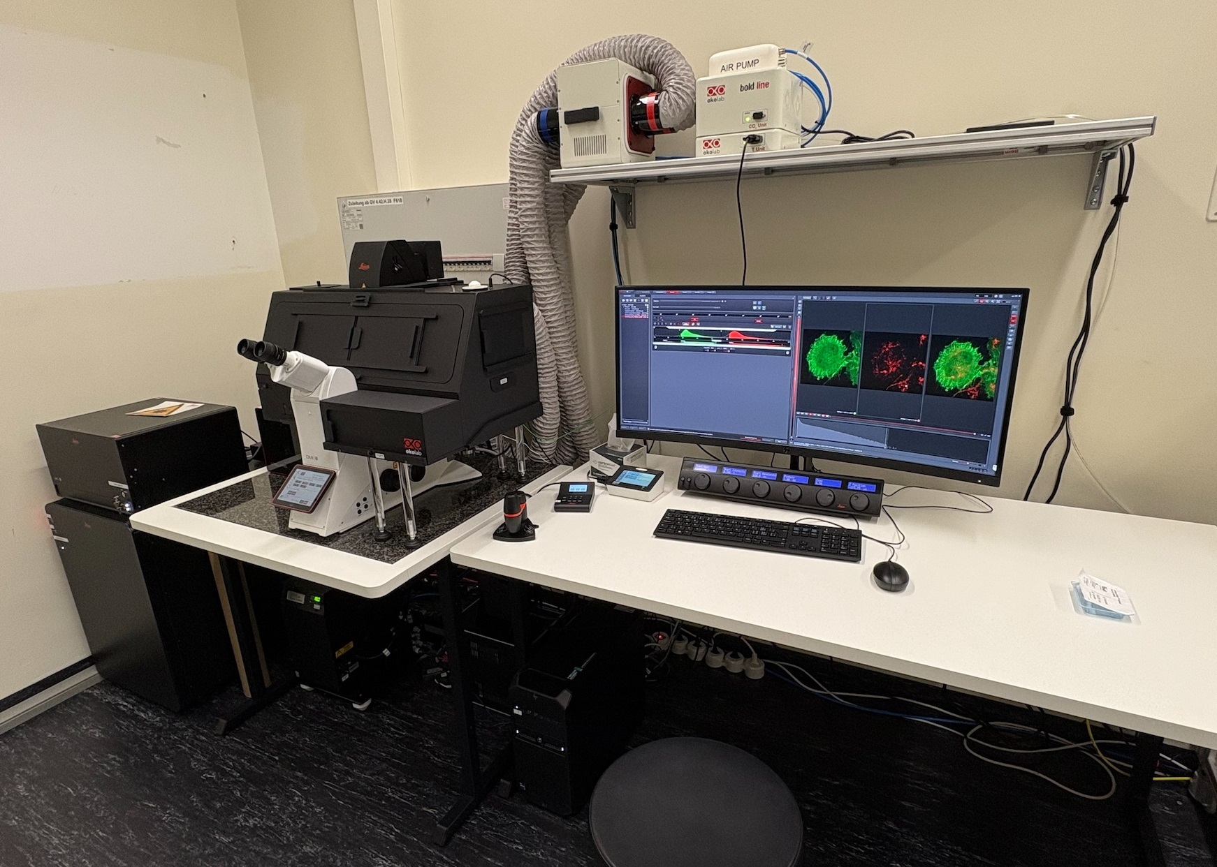
The Leica Stellaris is an automated confocal laser scanning microscope allowing simultaneous aquisition of 4 fluorescent channels as well as a transmission bright field channel. The SpectraPlex module provides a streamlined workflow to simplify panel creation, automate acquisition settings, and acquire data through advanced unmixing algorithms. The newest TauSense technology gives instant access to lifetime-based information, delivering additional insights to your experiments. In addition to the multicolor images in fixed samples the microscope can acquire large tile scans fully automated thanks to the LAS X Navigator and Navigator Expert. On top of that, this microscope also offers modules for FRET and FRAP advanced experiments.
Location
University Zurich, Irchel Campus, Room Y42-H-93.
Training Request
Follow this link to apply for an introduction to the microscope.
Technical Specifications
Light sources and lasers
- Halogen lamp for transmitted light
- External epi-fluorescence lamp - pE-300 white
- 405 nm diode laser
- Leica white light laser (WLL - 440 nm to 790 nm excitation)
Scanners
Dual scanning system:
Regular scanner: 1 - 2600 Hz
Resonant scanner: 8000 Hz
Objectives
| Name | Magnification | NA | Immersion | WD (mm) |
|---|---|---|---|---|
| HC PL FLUOTAR | 10x | 0.3 | Air | 11.0 |
| HC PL APO CS2 | 20x | 0.75 | Air | 0.62 |
| HC PL APO CS2 | 20x | 0.75 | IMM | 0.66 |
|
PL FLUOTAR |
40x | 1.00 | Oil |
0.08 |
| HC PL APO CS2 | 63x | 1.3 | Glycerol | 0.3 |
| HC PL APO CS2 | 63x | 1.4 | Oil | 0.14 |
CS2 objectives: improved color correction, perfect VIS-405; IMM = multi immersion (either water, glycerol or oil)
Detector System
5 PowerHybrid detectors:
- 3x HyD S providing unique flexibility and versatility for a wide range of confocal applications
- 1x HyD X
- 1x HyD R deliver optimized imaging in the near-infrared
One additional detector for transmission (Trans PMT). Excitation controlled by AOTF. Spectral detection.
Fluorescence Filters
| Name | Excitation Range | Excitation Filter | Dichroic | Emission Filter |
|---|---|---|---|---|
| DAPI | UV | BP 350/50 | 400 | BP 460/50 |
| GFP | blue | BP 470/40 | 495 | BP 525/50 |
| RHOD | green | BP 546/10 | 560 | BP 585/40 |
Make sure to acknowledge the Center for Microscopy in your publication to support us.
How to acknowledge contributions of the Center for Microscopy
Remarks
Literature and Links
Further information (internal UZH use only)
Follow this linkfor further background information, documents and links
Responsible Persons
If you have questions about the device please contact the responsible person.
Make sure to acknowledge the Center for Microscopy in your publication to support us.
How to acknowledge contributions of the Center for Microscopy