-

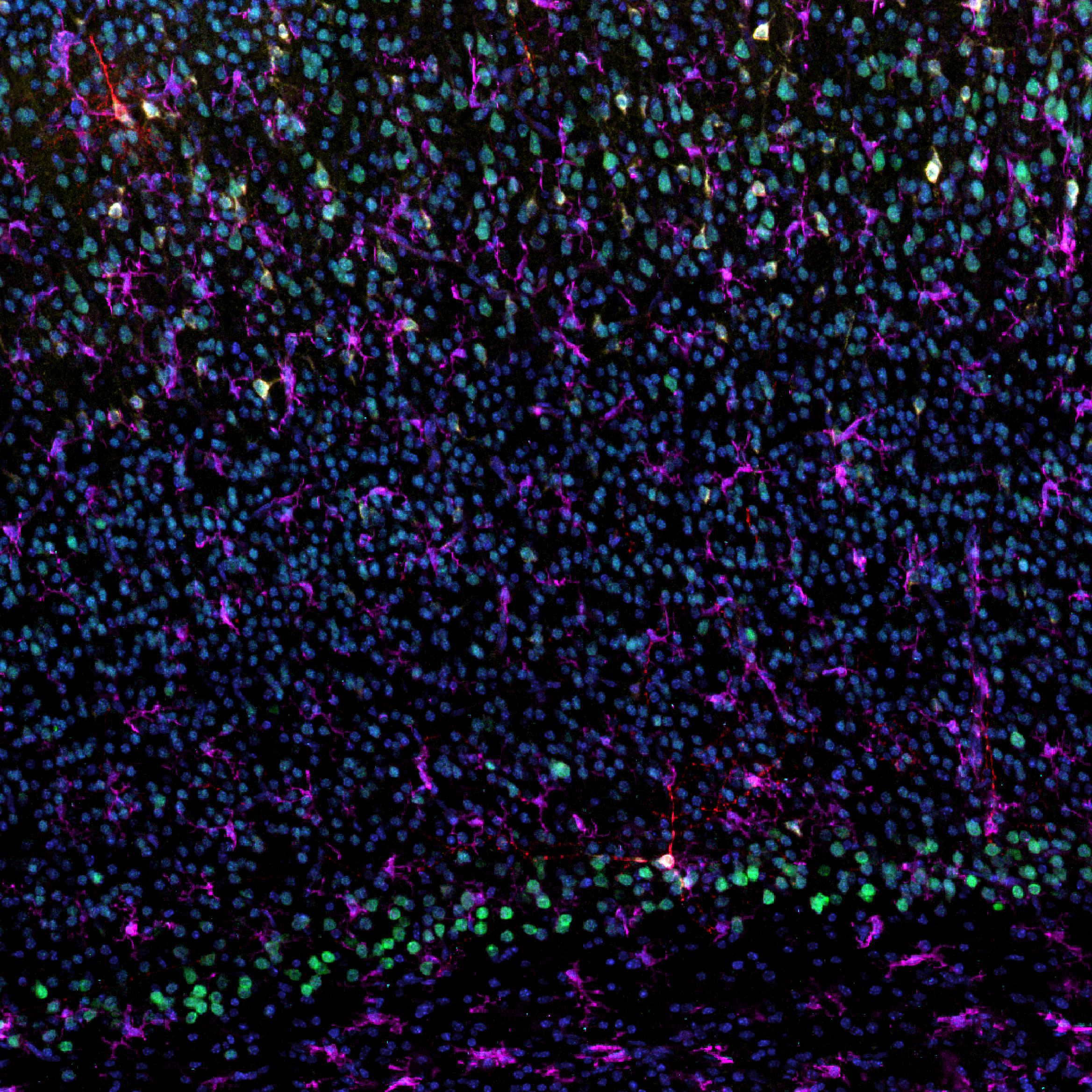
Welcome to the Center for Microscopy and Image Analysis (ZMB) at the University of Zurich – your partner in advanced biomedical imaging. As a leading academic core facility, we provide researchers from academia and industry with access to state-of-the-art light and electron microscopy technologies, complemented by expert scientific support and training. Our mission is to empower your research through deep technical expertise, tailored services and a collaborative spirit. Whether you're developing new imaging strategies, exploring complex biological systems or training the next generation of scientists, ZMB is here to support you at every step. Join a vibrant scientific community where innovation meets precision – and where your research can reach its highest potential through imaging.
-
.png "Thy1")
Welcome to the Center for Microscopy and Image Analysis (ZMB) at the University of Zurich – your partner in advanced biomedical imaging. As a leading academic core facility, we provide researchers from academia and industry with access to state-of-the-art light and electron microscopy technologies, complemented by expert scientific support and training. Our mission is to empower your research through deep technical expertise, tailored services and a collaborative spirit. Whether you're developing new imaging strategies, exploring complex biological systems or training the next generation of scientists, ZMB is here to support you at every step. Join a vibrant scientific community where innovation meets precision – and where your research can reach its highest potential through imaging.
-
.png "EB3")
Welcome to the Center for Microscopy and Image Analysis (ZMB) at the University of Zurich – your partner in advanced biomedical imaging. As a leading academic core facility, we provide researchers from academia and industry with access to state-of-the-art light and electron microscopy technologies, complemented by expert scientific support and training. Our mission is to empower your research through deep technical expertise, tailored services and a collaborative spirit. Whether you're developing new imaging strategies, exploring complex biological systems or training the next generation of scientists, ZMB is here to support you at every step. Join a vibrant scientific community where innovation meets precision – and where your research can reach its highest potential through imaging.
-
.png "mNeongreenactin")
Welcome to the Center for Microscopy and Image Analysis (ZMB) at the University of Zurich – your partner in advanced biomedical imaging. As a leading academic core facility, we provide researchers from academia and industry with access to state-of-the-art light and electron microscopy technologies, complemented by expert scientific support and training. Our mission is to empower your research through deep technical expertise, tailored services and a collaborative spirit. Whether you're developing new imaging strategies, exploring complex biological systems or training the next generation of scientists, ZMB is here to support you at every step. Join a vibrant scientific community where innovation meets precision – and where your research can reach its highest potential through imaging.
-
.png "Actin")
Welcome to the Center for Microscopy and Image Analysis (ZMB) at the University of Zurich – your partner in advanced biomedical imaging. As a leading academic core facility, we provide researchers from academia and industry with access to state-of-the-art light and electron microscopy technologies, complemented by expert scientific support and training. Our mission is to empower your research through deep technical expertise, tailored services and a collaborative spirit. Whether you're developing new imaging strategies, exploring complex biological systems or training the next generation of scientists, ZMB is here to support you at every step. Join a vibrant scientific community where innovation meets precision – and where your research can reach its highest potential through imaging.

.png "res")
.png)
.png)
.png)
.png)
.png)