Lightsheet Zeiss Lattice Lightsheet (Irchel)

ZEISS Lattice Lightsheet
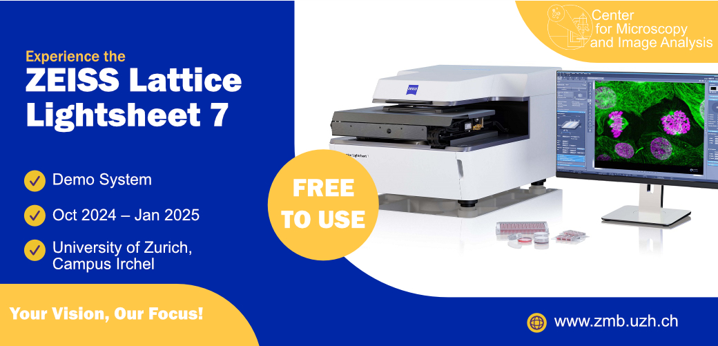
We are excited to offer the ZEISS Lattice Lightsheet 7 as a demo system, generously provided by ZEISS, free of charge for all trained users from mid-October 2024 to mid-January 2025.
The ZEISS Lattice Lightsheet 7 offers a fast and gentle light sheet fluorescence microscopy solution with an inverted configuration, designed for live and fixed sample imaging. The system is compatible with standard sample carriers featuring coverslip bottoms (#1.5), including 35 mm glass-bottom dishes, Labtek chambers, and IBIDI slides. Equipped with three laser lines (488 nm, 561 nm, and 640 nm), two Hamamatsu Fusion sCMOS cameras, and a 48x/1.0 NA detection objective, it enables subcellular resolution imaging with minimal photo damage. Fully incubated for live cell experiments, the system integrates acquisition and data processing through ZEN Blue, ensuring smooth workflows for extended imaging sessions.
Applications:
- Live cell imaging over extended periods
- High-volume imaging at fast speeds
- Single plane acquisition for high frame rates
Responsible Person |
|
Location |
University Zurich, Irchel Campus, Room Y42 H36. |
Training Request |
Follow this link to apply for an introduction to the microscope |
Light-sheet microscopy
|
|
Lattice-Light-sheet
|
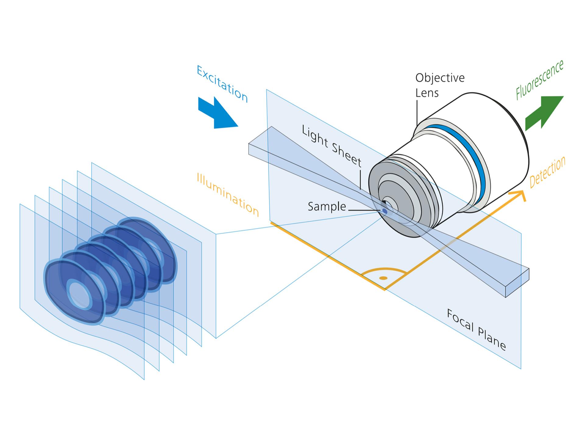
Lattice light sheet microscopy merges the benefits of traditional light sheet microscopy with near-isotropic resolution comparable to confocal imaging. By employing advanced beam-shaping technology, it generates lattice-shaped light sheets that are much thinner than conventional Gaussian light sheets, offering enhanced resolution without sacrificing imaging speed. This lattice pattern is formed using a Spatial Light Modulator (SLM) and is projected onto the sample after being refined by scanners, which dither the lattice to create a uniform, smooth light sheet for imaging. |
Technical Specifications
Microscope body |
|
||||||||||
Light Sources and Lasers |
|
||||||||||
Camera System |
|
||||||||||
Environmental control |
|
||||||||||
Accessories |
|
||||||||||
Available Optics |
|
||||||||||
Links & Literature |
Make sure to acknowledge the Center for Microscopy and Image Analysis in your publication to support us.
How to acknowledge contributions of the Center for Microscopy and Image Analysis
