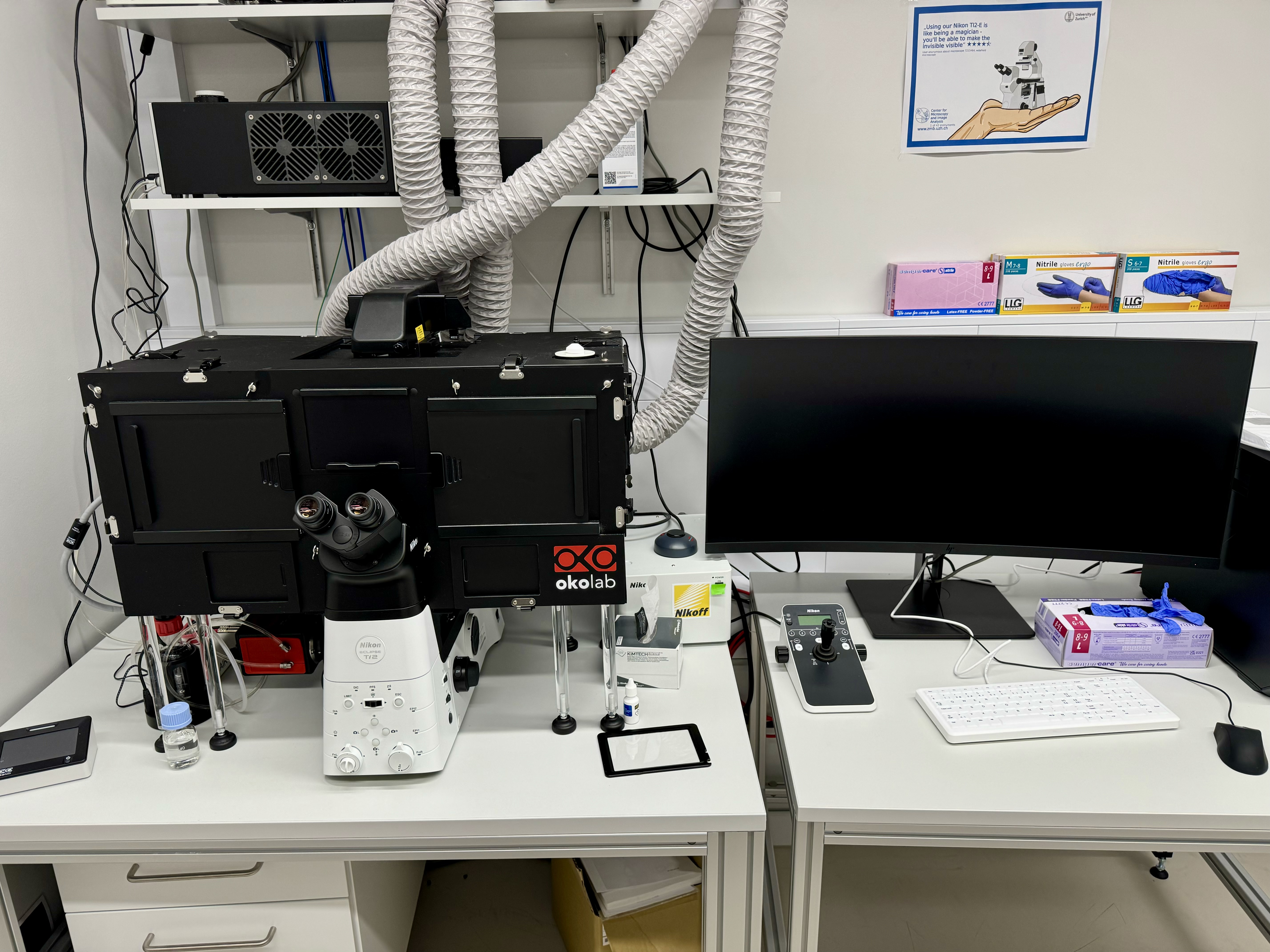
Wide-field Nikon-Ti2-E (Irchel)

Nikon Ti2-E
This inverted wildfield system is suited to image live cells as well as fixed samples in fluorescence and transmission light modes. A temperature controlled environment is possible for live cell experiments. The system does not have CO2 control. It is well suited for phase contrast imaging of bacteria and does allow complex imaging tasks.
Responsible Person |
|
Location |
University Zurich, Irchel Campus, Room Y38-M-77. |
Training Request |
Follow this link to apply for an introduction to the microscope |
The Nikon Ti2-E features the following modalities:
Wide-field microscopy |
Fluorescence wide-field microscopy is a technique where the entire specimen is illuminated uniformly, causing fluorophores within the sample to emit light. This emitted fluorescence is then captured by a camera or detector, producing an image that shows the distribution of fluorescent molecules. It's widely used for observing cellular structures, proteins, and other biomolecules tagged with fluorescent markers. The technique offers high sensitivity and allows for real-time imaging of live cells, making it invaluable in biological and medical research. |
Differential interference contrast microscopy (DIC) |
Differential interference contrast (DIC) microscopy is an advanced optical microscopy technique that enhances the contrast in unstained, transparent specimens. DIC utilizes polarized light split into two beams that pass through the specimen at slightly different angles. When these beams recombine, the differences in optical path length cause interference, creating an image with high contrast and pseudo-3D effect. This technique is particularly useful for observing fine details and structures within live cells and tissues without the need for staining, making it ideal for dynamic studies in cell biology. |
Phase contrast microscopy (PH) |
Phase contrast microscopy is a technique designed to enhance the contrast of transparent and colorless specimens, such as living cells, by converting phase shifts in light passing through the specimen into changes in intensity. It employs a phase plate to create constructive and destructive interference, which highlights differences in refractive index within the sample. This allows for the detailed visualization of cellular structures and organelles that are otherwise invisible under standard bright-field illumination. Phase contrast microscopy is widely used in biological research for examining live, unstained cells and tissues. |
Technical Specifications
Microscope body |
|
|||||||||||||||||||||||||||||||||||||||||||||||||||||||||||||||||||||||||||||||||||||||||||||||
Light Sources |
|
|||||||||||||||||||||||||||||||||||||||||||||||||||||||||||||||||||||||||||||||||||||||||||||||
Camera System |
|
|||||||||||||||||||||||||||||||||||||||||||||||||||||||||||||||||||||||||||||||||||||||||||||||
Environmental control |
|
|||||||||||||||||||||||||||||||||||||||||||||||||||||||||||||||||||||||||||||||||||||||||||||||
Accessories |
|
|||||||||||||||||||||||||||||||||||||||||||||||||||||||||||||||||||||||||||||||||||||||||||||||
Available Optics |
PH = phase contrast, DIC = Differential Interference Contrast |
|||||||||||||||||||||||||||||||||||||||||||||||||||||||||||||||||||||||||||||||||||||||||||||||
Available Filters |
|
|||||||||||||||||||||||||||||||||||||||||||||||||||||||||||||||||||||||||||||||||||||||||||||||
User Guide |
will come soon! | |||||||||||||||||||||||||||||||||||||||||||||||||||||||||||||||||||||||||||||||||||||||||||||||
Links & Literature |
||||||||||||||||||||||||||||||||||||||||||||||||||||||||||||||||||||||||||||||||||||||||||||||||
Make sure to acknowledge the Center for Microscopy and Image Analysis in your publication to support us.
How to acknowledge contributions of the Center for Microscopy and Image Analysis