Lunaphore COMET (Schlieren)
Lunaphore COMET
Make sure to acknowledge the Center for Microscopy and Image Analysis in your publication to support us.
How to acknowledge contributions of the Center for Microscopy and Image Analysis
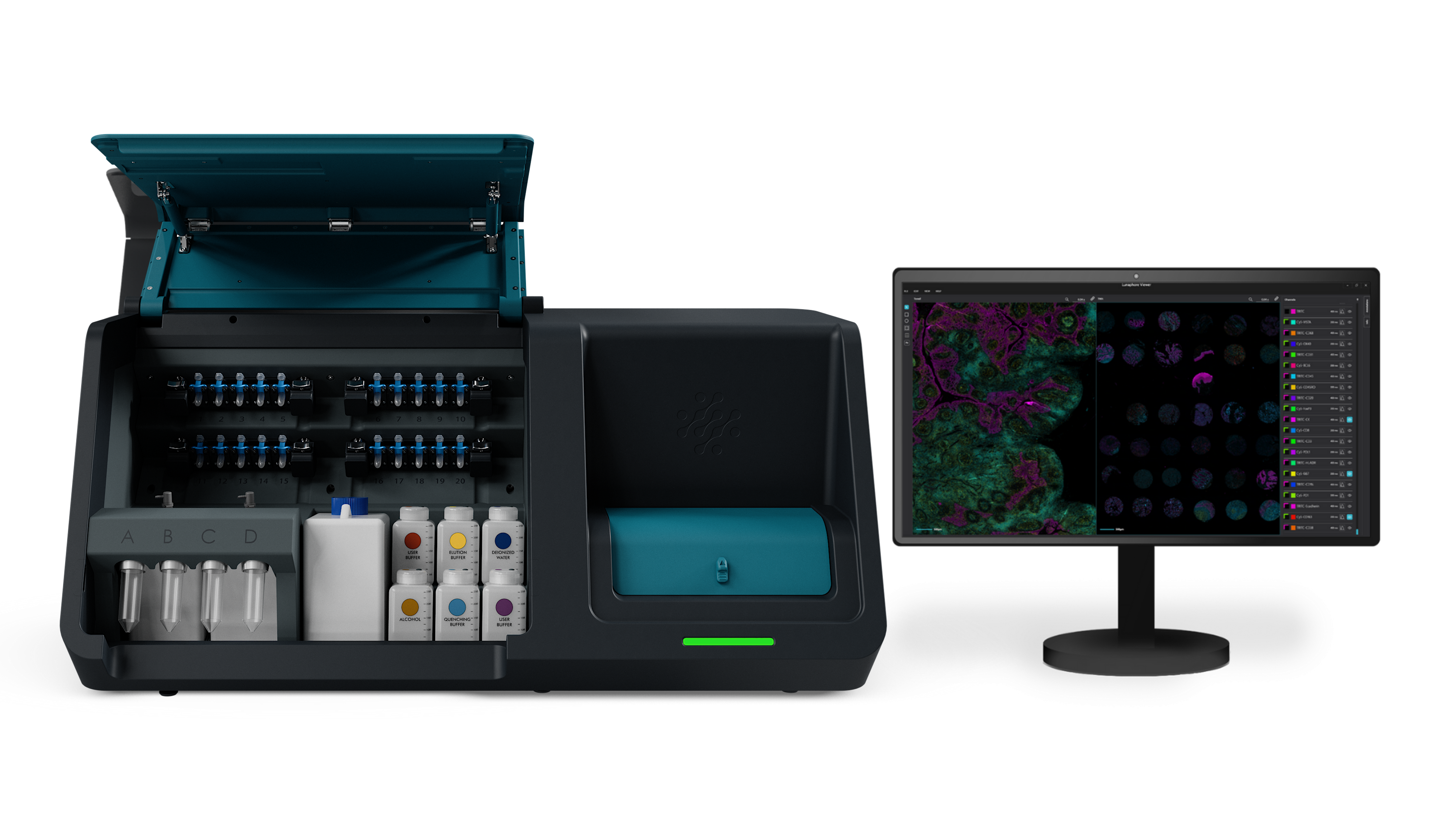
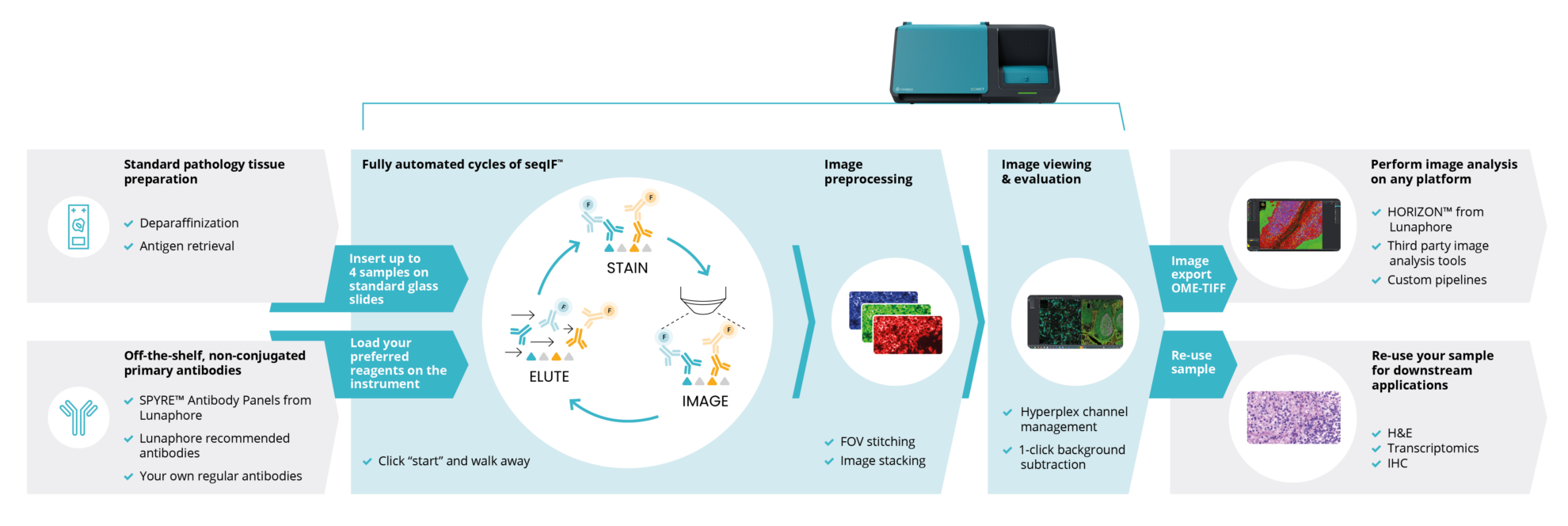
The Lunaphore COMET system is a fully automated, high-throughput multiplex immunofluorescence platform designed for spatial biology and tissue profiling. By integrating microfluidics, staining, and imaging in one streamlined workflow, COMET enables the analysis of multiple biomarkers (40+ plex) on a single tissue section without compromising sample integrity.
Responsible Person |
|
Location |
University Zurich, Schlieren Campus, Room WAD K134(532) |
Training Request |
Follow this link to apply for an introduction to the microscope |
Applications |
Multiplex sequential immunofluorescence (seqIF) and RNAscope HiPlex |
| Plex-level |
|
Technical Specifications
Slide capacity |
4 slides |
|
Slide compatibility |
Standard histology slides - 75 mm x 25 mm (3 in x 1 in), 1 mm thick, positively charged such as Epredia SuperFrost Plus |
|
Sample fixation |
Optimized for FFPE samples and frozen sections | |
Sample thickness |
3-10 μm | |
Automation level |
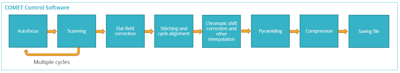
Fully automated: staining, target probe hybridization, image acquisition and image preprocessing | |
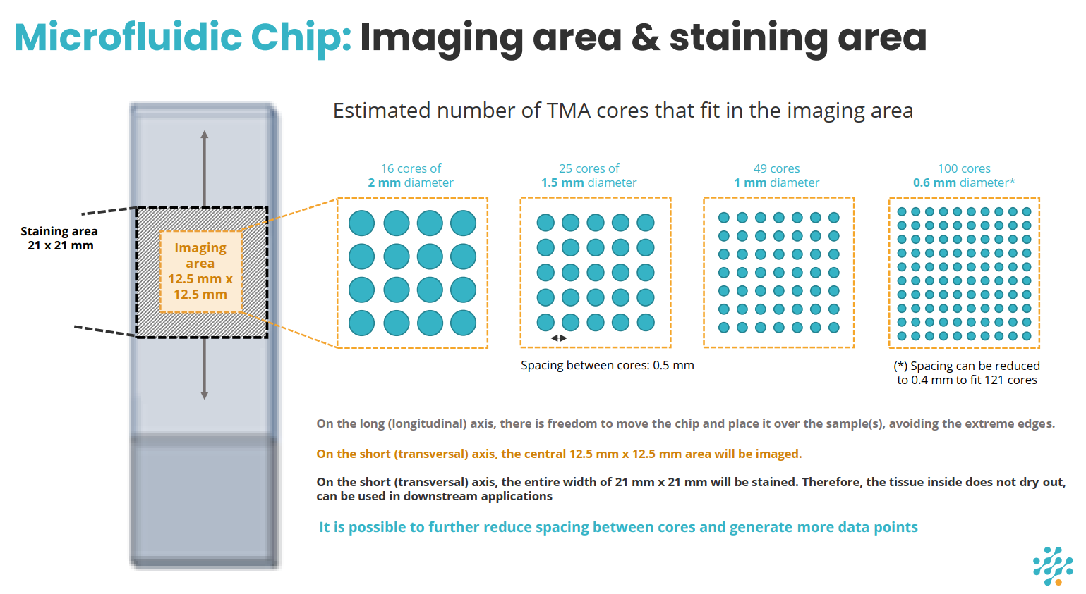
Maximum imaging area |
12.5 mm x 12.5 mm | |
Maximum staining area |
21 mm x 21 mm | |
|
|
||
Throughput |
Measured for a 9 mm x 9 mm imaging area: 20 slides/week for 20-plex protein panel ; 12 slides/week for 40-plex protein panel | |
Reagents |
Open choice of primary antibodies (non-conjugated): |
|
| Secondary antibodies: |
|
|
Software |
|
|
Available Optics |
20X / 0.7 NA | |
Image resolution |
0.28 µm/pixel | |
Available Channels |
DAPI, FITC, TRITC, Cy5, Cy7 | |
Output file format |
OME-TIFF | |
|
|
||
User Guide |
COMET USER Guide | |
Links & Literature |
||
Reagents
|
Application |
Reagent/Item |
Cat. No. |
|
RNAscope HiPlex Pro for COMET 12-plex Kit |
RNAscope HiPlex Pro AMP 1 |
ACD (Bio-Techne) 322075 |
|
RNAscope HiPlex Pro AMP 2 |
||
|
RNAscope HiPlex Pro AMP 3 |
||
|
RNAscope HiPlex Pro Fluoro T1-T4 |
||
|
RNAscope HiPlex Pro Fluoro T5-T8 |
||
|
RNAscope HiPlex Pro Fluoro T9-T12 |
||
|
RNAscope HiPlex PretreatPro™ |
||
|
RNAscope HiPlex Pro Probe Diluent |
||
|
RNAscope HiPlex Pro DAPI (1 mg/mL) |
||
|
RNAscope HiPlex Cleaving Stock Solution v2 |
||
|
RNAscope HiPlex Pro Imaging Buffer Part A |
||
|
RNAscope HiPlex Pro Imaging Buffer Part B |
||
|
|
||
|
RNAscope HiPlex Pro for COMET 12-plex Kit |
RNAscope HiPlex Pro AMP 1 |
ACD (Bio-Techne) 322075 |
|
RNAscope HiPlex Pro AMP 2 |
||
|
RNAscope HiPlex Pro AMP 3 |
||
|
RNAscope HiPlex Pro Fluoro T1-T4 |
||
|
RNAscope HiPlex PretreatPro™ |
||
|
RNAscope HiPlex Pro Probe Diluent |
||
|
RNAscope HiPlex Pro DAPI (1 mg/mL) |
||
|
RNAscope HiPlex Cleaving Stock Solution v2 |
||
|
RNAscope HiPlex Pro Imaging Buffer Part A |
||
|
RNAscope HiPlex Pro Imaging Buffer Part B |
||
|
|
||
|
Control Probes |
RNAscope HiPlex12 CS Negative Control Probe |
ACD (Bio-Techne) 324347 |
|
RNAscope HiPlex12 CS Positive Control Probe – Hs |
ACD (Bio-Techne) 324317 |
|
|
RNAscope HiPlex12 CS Positive Control Probe – Mm |
ACD (Bio-Techne) 324437 |
|
|
RNAscope HiPlex12 CS Positive Control Probe – Rn |
ACD (Bio-Techne) 324337 |
|
|
|
||
|
Extras |
RNAscope Control Slide -Human Hela Cell Pellet |
ACD (Bio-Techne) 310045 |
|
RNAscope HiPlex Cleaving Stock Solution v2 |
ACD (Bio-Techne) 324399 |
|
|
|
||
|
Consumables |
COMET chip (20-pack) |
Lunaphore MK03 |
|
Multistaining Buffer 20x |
Lunaphore BU06 |
|
|
Dewax and HIER Buffer L (pH 6) |
Epredia AR02 |
|
|
Dewax and HIER Buffer H, pH 9 |
Epredia AR03 |
|
|
Imaging Buffer |
Lunaphore BU09 |
|
|
Elution Buffer Kit |
Lunaphore BU07-L |
|
|
Blocking Buffer |
Lunaphore BU10 |
|
|
Quenching Buffer Kit |
Lunaphore BU08-L |
|
|
20X SSC |
Thermo Fisher 15557044 |
|
|
Xylene (or xylene-alternative, e.g. Histoclear) |
Merck H2779 |
|
|
50 mL conical tubes |
Falcon 352070 |
|
|
2 mL microtubes |
Eppendorf 0030123344 |
|
|
SuperFrost Plus Adhesion Slides |
Epredia J1830AMNZ |
|
|
Alexa Fluor™ Plus 488 Goat anti-Mouse IgG |
Thermo Fisher A32723 |
|
|
Alexa Fluor™ Plus 488 Goat anti-Rabbit IgG |
Thermo Fisher A32731 |
|
|
Alexa Fluor™ Plus 488 Goat anti-Rat IgG |
Thermo Fisher A48262 |
|
|
Alexa Fluor™ Plus 555 Goat anti-Mouse IgG |
Lunaphore DR555MS |
|
|
Alexa Fluor™ Plus 647 Goat anti-Mouse IgG |
Lunaphore DR647MS |
|
|
Alexa Fluor™ Plus 555 Goat anti-Rabbit IgG |
Lunaphore DR555RB |
|
|
Alexa Fluor™ Plus 647 Goat anti-Rabbit IgG |
Lunaphore DR555RB |
|
|
Alexa Fluor™ Plus 555 Goat anti-Rat IgG |
Lunaphore DR555RT |
|
|
Alexa Fluor™ Plus 647 Goat anti-Rat IgG |
Lunaphore DR647RT |
|
|
Alexa Fluor™ 750 Goat anti-Rabbit IgG |
Thermo Fisher A-21039 |
|
|
Alexa Fluor™ 750 Goat anti-Mouse IgG |
Thermo Fisher A-21037 |
|
|
DAPI solution |
Lunaphore DR100 |
|
|
|
||
|
SPYRE™ Immuno-Oncology Core Panel Kit, human |
This panel includes 13 validated monoclonal antibodies for FFPE human tissues: αSMA, CD3, CD4, CD8, CD11c, CD20, CD45, CD56, CD68, FOXP3, Ki-67, PD-1, PD-L1
|
Lunaphore MP10010HU |